High-resolution 3D mapping of the human hypothalamus in 10 postmortem brains
Alexey Chervonnyy – Hector Fellow Katrin Amunts
Our study aims to analyse and map the cytoarchitecture of the human hypothalamus in histological sections of 10 postmortem brains. As a result, we want to develop a high-resolution 3D reconstructed histological model of the hypothalamus and its nuclei as a tool for assessing the structure-function relationship and a probabilistic cytoarchitectonic map of the hypothalamus that will reflect the variability of hypothalamic nuclei between individual brains, in terms of size and location in standard reference space.
The hypothalamus is part of the diencephalon that plays a key role in maintaining homeostasis of neuroendocrine, behavioural and autonomic processes. It consists of several nuclei, which differ in their microstructure, connectivity and function.
Studying structure-functional relationships of the hypothalamus is becoming more feasible with increasing spatial resolution of modern neuroimaging in the living human brain. However, it is difficult to localize brain activations in the functionally different nuclei, which are hardly visible in routine MR. Currently, there are no maps, which inform neuroimaging studies about the microstructural segregation of the hypothalamus in 3D space.
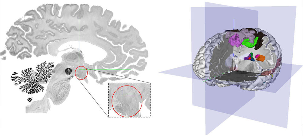
We therefore aim to develop a cytoarchitectonic map of the hypothalamus in the BigBrain – a high-resolution 3D-reconstructed histological model of the human brain, using a deep-learning based mapping tool. The resulting map will capture the shape and extent of the hypothalamic nuclei with cellular precision. Thus, the map will bridge the microscopic structure of the hypothalamus and MR-based studies at the macroscale.
Also, we will develop probabilistic cytoarchitectonic maps of the hypothalamus. Therefore, we will study serial histological sections of 10 postmortem brains and map the nuclei over their full extent. Maps will be transferred to standard references space to link them with neuroimaging data, to serve as an anatomical reference for studies of healthy brains and patients. They will be freely available to the scientific public.
Sagittal section of the human brain with the region of interest marked in the red circle (left); 3D image of the human brain from the Big Brain dataset (right).

Alexey Chervonnyy
Heinrich Heine Universität DüsseldorfSupervised by

Katrin Amunts
Medicine & Brain Research

